
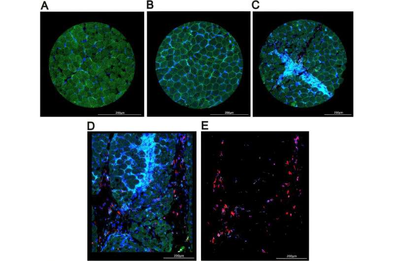
Immunofluorescent imaging of the muscle biopsy regions of interest (ROIs). (A-E) Representative fluorescent images of ROI types selected for sequence analysis and associated staining markers: (A) control muscle; (B) JDM muscle ROI with no infiltrating immune cells; (C) JDM muscle enriched with immune cells (CD45+); (D) CD68+ macrophage-enriched region. (E) Segmented CD68+ cells in the same region shown in (D). Staining antibodies: laminin (green), DNA nuclear stain (blue), CD68 (red), and CD45 (cyan), indicating muscle fibers, nuclei, macrophages, and leukocytes, respectively. (B-E) are derived from patient JDM3. JDM, juvenile dermatomyositis. Credit: Annals of the Rheumatic Diseases (2025). DOI: 10.1016/j.ard.2025.07.015
Children with a rare, debilitating muscle disease could benefit from the findings of new research by experts at UCL and Great Ormond Street Hospital (GOSH).
For the study, published in Annals of the Rheumatic Diseases, the scientists closely analyzed muscle samples from three children with juvenile dermatomyositis (JDM) and discovered the mitochondria, small energy-producing structures inside cells, were not functioning correctly.
It is hoped that this research will enable future treatments to be identified, especially for children who do not respond well to current treatment.
William Magee, father of one of the children who took part in the study, welcomed the breakthrough, saying that it "opens the doors to a whole new type of treatment."
Juvenile dermatomyositis
JDM is a rare condition that causes muscle weakness and skin rashes. It inflames the capillaries—the body's tiny blood vessels—causing a rash on parts of the body like fingers and elbows. In some cases, the disease causes calcium deposits under the skin or deeper in tissues, which can restrict the movement of the joints.
It often causes problems with muscles, which can make everyday activities like walking or playing very difficult. JDM can also cause problems with other organs, like lungs or intestines, and in turn lead to organ damage.
There are two to four new cases per million children a year. However, this is incidence (new cases); the actual prevalence of the disease is hard to estimate.
Under the microscope
Researchers were able to closely analyze muscle samples from children with JDM and compare them to healthy muscles. They used a special new technology called spatial transcriptomics, which is like a super-powered microscope that can identify which genes are active in tiny parts of the muscle. This helped researchers understand what was happening inside the muscles of children with JDM.
They identified two differences in the muscles of children with the condition. The first was mitochondrial dysfunction. Researchers found that the mitochondria in the cells of children with JDM were not functioning correctly, even in muscles that didn't seem weak.
The second difference they found was that the body's immune system goes into overdrive, through a process known as interferon activation. Essentially, the immune system tries to fight off an infection that isn't there, which can make children feel weak. While this process is already well known in JDM, this study was the first time it's been mapped in such detail directly within the muscle.
Interestingly, the scientists found that these two problems don't always occur together.
Senior author and consultant pediatrician Professor Lucy Wedderburn (UCL Great Ormond Street Institute of Child Health) said, "While more research into JDM is needed, this is a big step forward. It shows that looking at tiny details in muscle tissue can reveal important clues—and might help us find better ways to treat JDM in the future, for example by focusing on fixing the mitochondria, not just calming the immune system. This is an important finding as current treatments mostly focus on reducing inflammation."
Lucia's story
Five-year-old twin Lucia, from Twickenham in southwest London, was diagnosed with JDM in August 2024. However, her identical twin sister Isabella does not have the condition.
Lucia's parents, William and Caitriona, noticed something wasn't quite right when she had a red rash on the joints on her fingers and toes during a trip to New Zealand at Christmas 2023.
They thought it might be related to cold weather, but when it got worse despite the warm weather, they began to grow concerned.
On returning to the UK, they went to see their GP and began the process of seeing several specialists until they were seen by now-retired GOSH doctor Clarissa Pilkington, who had set up a specialist unit for conditions like JDM at the hospital.
Lucia was diagnosed with JDM and started on two treatments in August 2024, a steroid and an immune-suppressing treatment called methotrexate.
Dad William Magee, a management consultant, said, "We were pleased to have an early diagnosis which was crucial to Lucia starting treatment quickly. However, the steroids weren't as effective as hoped, so Lucia was moved onto intravenous immunoglobulin, which involves being admitted to hospital for two days each month for a transfusion of antibodies to boost her immune system.
"This research suggesting the link to mitochondria is so important because it opens the doors to a whole new type of treatment.
"It's very reassuring to know that it could lead to treatments that address the underlying causes, not just the symptoms of the disease—it is common for children with JDM to go into remission and then have flareups in the future. We'd like Lucia and children with this condition to spend as little time as possible in hospital, so any research which leads to potentially more effective treatments can only be a good thing."
He praised the skill of the GOSH clinicians, adding the treatment Lucia had received had been excellent. He said, "We were lucky—the doctors caught Lucia's condition early. Not everyone with this condition is so fortunate."
Study co-lead Dr. Merry Wilkinson (UCL Great Ormond Street Institute of Child Health), said, "This new and exciting research has shown us that there are mitochondrial problems in the muscle of JDM patients.
"We now need to investigate in the lab the use of existing drugs or developing new ones to improve mitochondrial health in JDM. There is a dire need for better treatments with fewer side effects and hopefully this research will start to address this problem.
"We are extremely privileged to work on the Juvenile Dermatomyositis Cohort and Biobank study, one of the largest databases of both clinical and biological JDM samples in the world, and very grateful to all the patients and families who donate samples to research like ours."
Patients and families were involved at every stage of this research, including study conception, delivery and analysis through patient and public involvement and engagement group.
More information: Aris E. Syntakas et al, Spatial transcriptomic analysis of muscle biopsy from patients with treatment-naive juvenile dermatomyositis reveals mitochondrial abnormalities despite disease-related interferon-driven signature, Annals of the Rheumatic Diseases (2025). DOI: 10.1016/j.ard.2025.07.015 Journal information: Annals of the Rheumatic Diseases


Post comments