
byIngrid Fadelli, Medical Xpress
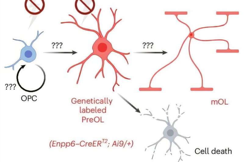
Schematic showing the potential effect of neuronal activity on oligodendrogenesis, acting through its influence on both OPCs and preOLs. Credit:Nature Neuroscience(2025). DOI: 10.1038/s41593-025-02110-1.
Nerve fibers in the brain and spinal cord are wrapped in an insulating sheath known as myelin. For a long time, this barrier, which is essentially the brain's white matter, was believed to serve the main function of speeding up the propagation of electrical signals along axons (i.e., the extensions via which neurons communicate).
This is because when myelin is damaged or lost due to strokes, immune disorders, infections, metabolic disorders or some other conditions, the transmission of electrical signals slows down. This can prompt the emergence of various symptoms, such as changes in vision, motor control issues, fatigue and sensory changes.
Myelin is known to be created by cells known as oligodendrocytes, which originate from the division of so-called oligodendrocyte precursor cells (OPCs). In diseases associated with a loss of myelin, OPCs are known to be unable to form mature myelin, with pre-myelinating oligodendrocytes often observed near demyelinated lesions.
Researchers at University of Texas Southwestern Medical Center recently genetically engineered a new type of mice that is better suited for mapping pre-myelinating oligodendrocytes and studying their responses to the activity of neurons.
This valuable tool, introduced in a paper published inNature Neuroscience, could help to shed new light on the processes underpinning the formation of myelin and its loss or degradation in various diseases.
"Recent work has shown that myelin does far more than speed up electrical signal propagation: it provides metabolic and trophic support to axons, responds dynamically to neuronal activity by adjusting insulation levels, and is among the earliest cell types affected in neurodegenerative diseases such as Alzheimer's," Aksheev Bhambri, first author of the paper, told Medical Xpress.
"Understanding how myelin develops is therefore crucial. The paper came about from my postdoc mentor Dr. Sun's dream of having a mouse line to target the pre-myelinating oligodendrocytes. Pre-myelinating oligodendrocytes are basically the overlooked 'middle child' of myelination."
Pre-myelination is a crucial stage during which OPCs differentiate to the pre-myelinating stage, subsequently maturing to form myelin, while it is decided what axon or specific part of axons they will myelinate. Notably, many of these cells never become mature enough to form myelin and die before then.
"Dr. Sun attempted to generate a mouse line that could be used to study pre-myelinating oligodendrocytes twice during his postdoc at Stanford, but without success," said Bhambri. "When he shared the idea with me, I was immediately fascinated by the potential value of such a tool for the myelin field."
The main objective of this recent work was to develop a new resource that would allow neuroscientists to reliably visualize and study premyelinating oligodendrocytes in vivo, specifically in living mice. In collaboration with Dr. Sharma at the Children's Research Institute mouse engineering department, Bhambri and Dr. Sun were ultimately able to create the Enpp6-CreERT2 mouse line.
"While we were developing this mouse line, several influential studies onadaptive myelinationwere emerging, showing that neuronal activity can modify myelination, however, all focused on communication between neurons and oligodendrocyte precursor cells (OPCs)," said Bhambri.
"We wondered whether pre-myelinating oligodendrocytes, the cells that make decisions about where to myelinate, might also respond to neuronal activity. Realizing that our new mouse line offered a unique way to test this question was a true 'eureka moment.'"
In addition to developing a new resource to study pre-myelination processes, the researchers thus wished to assess its potential for probing the responses of pre-myelinating oligodendrocytes to neuronal activity.
The mouse line created by the researchers is characterized by the introduction of the CreERT2 enzyme into a gene that is active in pre-myelinating oligodendrocytes.
As part of their study, Bhambri and his colleagues showed that they could permanently label these cells in the mice and then track their changes over time.
"We studied pre-myelinating oligodendrocytes which are generated when oligodendrocyte precursor cells differentiate," explained Bhambri.
"After generating the Enpp6-CreERT2mouse line, we crossed it with a reporter mouse carrying a tdTomato fluorescent gene. In these reporter mice, a stop codon blocks tdTomato expression until it is removed by Cre recombinase. Because our mouse line expresses a tamoxifen-inducible form of Cre (CreERT2), administering tamoxifen activates Cre specifically in pre-myelinating oligodendrocytes, allowing tdTomato expression and enabling us to visualize these cells."
The researchers subsequently examined the brains of the engineered pups, using an approach known as immunohistochemistry to detect tdTomato-positive cells. With this technique and other genetic tools, they confirmed that the cells they labeled were indeed pre-myelinating oligodendrocytes.
"We did this by combining immunohistochemistry with in-situ hybridization for stage-specific RNA markers," said Bhambri. "To ensure that our tool labeled only pre-myelinating oligodendrocytes, we carried out a comprehensive characterization using immunohistochemistry, electrophysiology, and single-cell RNA sequencing."
To study how neuronal activity affected pre-myelinating oligodendrocytes, Bhambri and his colleagues carried out a behavioral experiment in which they trimmed the whiskers of newly born mice pups, which reduced the sensory input that their brain, specifically the somatosensory cortex, received.
"Using our reporter line, we then examined how decreased neuronal activity impacted pre-myelinating oligodendrocytes," said Bhambri.
"We found that reduced activity diminished both their survival and maturation. In-situ hybridization further revealed a decrease in the expression of neurotransmitter receptors under low-activity conditions, suggesting a mechanistic link between neuronal activity and pre-myelinating oligodendrocyte development."
This recent study introduces a new valuable resource that could soon be used by neuroscientists worldwide to study pre-myelinating oligodendrocytes, as well as de-myelination processes associated with specific diseases.
The single-cell dataset that Bhambri and his colleagues compiled when examining these cells in their genetically engineered mice is publicly available and can beaccessed by other research teams via a user-friendly web interface.
"We discovered that a small proportion of pre-myelinating oligodendrocytes can survive, without maturing, for up to 8 days in young neonates but not in older animals," said Bhambri. "Whether this is linked to the immature neuronal circuits still present in young animals as compared to old is an open question. We also observed that neuronal activity can regulate pre-myelinating oligodendrocyte survival and maturation."
The team's initial observations could have important implications for the study of various diseases, including multiple sclerosis (MS) and preterm white matter injury (pWMI). In these diseases, pre-myelinating oligodendrocytes fail to effectively mature and make myelin.
Future work could build on the team's findings to devise new interventions aimed at promoting the maturation of these cells. This might be achieved, for instance, by increasing neuronal activity via deep brain stimulation or anti-depressants, such as selective serotonin re-uptake inhibitors (SSRIs).
"The Sun Lab is now using the Enpp6-CreERT2 mouse line to identify the molecular mechanisms that regulate premyelinating oligodendrocyte survival in diseases such as MS and pWMI," said Bhambri.
"By discovering pathways that enhance remyelination, we hope to move closer to developing new therapeutic strategies. We also aim to generate additional tools to address deeper, unanswered questions in myelin biology, particularly how axons regulate myelin development beyond classical neurotransmitter-based signaling."
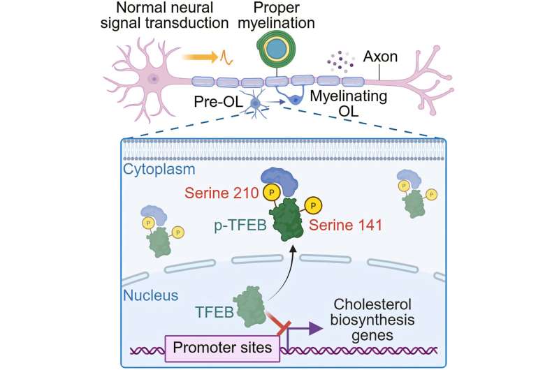
Graphical abstract. Credit:Cell Reports(2025). DOI: 10.1016/j.celrep.2025.116252
Other researchers in Dr. Lu Sun's laboratory at UT Southwestern recently published a new paper inCell Reports, where they presented the results of a study led by graduate student Dr. Yihe Zhang.
As part of this study, Dr. Zhang generated several new mouse lines that are more promising for probing how axons regulate the production of myelin.
© 2025 Science X Network
More information Aksheev Bhambri et al, Genetic targeting of premyelinating oligodendrocytes reveals activity-dependent myelination mechanisms, Nature Neuroscience (2025). DOI: 10.1038/s41593-025-02110-1 . Yihe Zhang et al, Neurons sequester the cholesterol-inhibiting TFEB in oligodendrocyte cytoplasm to safeguard myelination and neural function, Cell Reports (2025). DOI: 10.1016/j.celrep.2025.116252 Journal information: Cell Reports , Nature Neuroscience


Post comments