
byUT Southwestern Medical Center
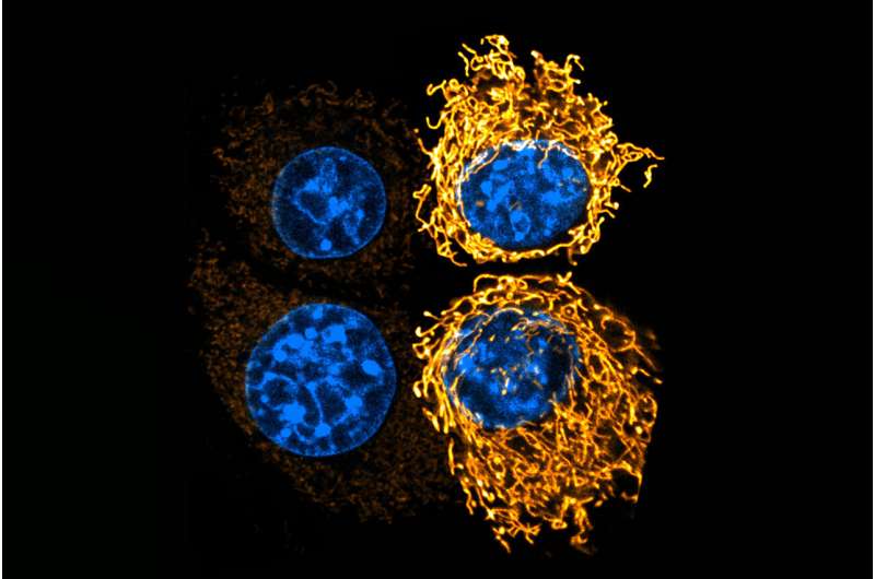
Credit:Cancer Cell(2025). DOI: 10.1016/j.ccell.2025.09.001
UT Southwestern Medical Center researchers have identified two distinct populations of cells known as antigen-presenting cancer-associated fibroblasts (apCAFs) that appear to support the survival and growth of malignant tumors. Their findings,reportedinCancer Cell, could one day lead to new therapies for notoriously hard-to-treat cancers, including pancreatic cancer and advanced colorectal cancer (CRC) that has spread throughout the abdomen, known as peritoneal metastasis.
"We have uncovered two groups of fibroblasts integrated within tumors and defined their spatial microenvironment that control the mechanism of cancer growth, metastasis, andtreatment resistance, suggesting a new strategy to therapeutically target atumor's supportive tissue," said Huocong Huang, M.D., Ph.D., Assistant Professor of Surgery and Immunology at UT Southwestern.
Dr. Huang co-led the study with Alex Kim, M.D., Ph.D., Assistant Professor of Surgery. Both are members of the Harold C. Simmons Comprehensive Cancer Center at UT Southwestern.
Over the past two decades, researchers have had a growing understanding that cancerous tumors aren't made only of cancer cells but instead are a heterogeneous mixture of many cell types. These include fibroblasts, which play vital roles in preserving tissue integrity, regulating inflammatory response, and facilitating wound repair. Although scientists initially thought that all cancer-associated fibroblasts (CAFs) were the same, advances in genetic profiling have shown that there are three subtypes.
In 2022, Dr. Huang's team was among the researchers who discovered one of these subtypes, now known as apCAFs, in a mouse model ofpancreatic cancer. Their findings showed that these cells express immune molecules on their surfaces and appear to regulate the activity of immune cells—called T cells—in tumors. However, little else was known about these cells, including whether they exist in human cancers, their origins in different cancer types, and where they tend to reside within tumors.
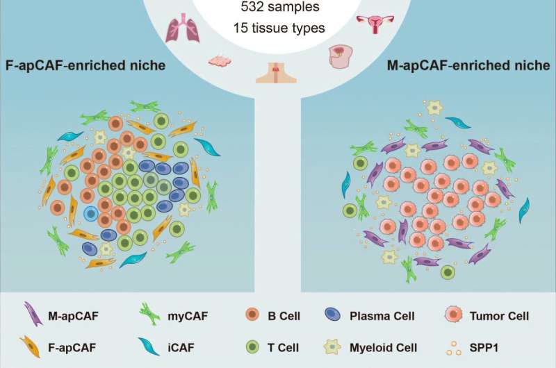
To learn more, Dr. Huang, Dr. Kim, and their UTSW colleagues combined information from multiple existing datasets of human cancer single-cell RNA sequencing (scRNA-seq). The technique allows researchers to analyze gene activity at the level of individual cells, making it possible to distinguish different cell types. Using this wealth of data—generated from more than 2.5 million cells in 532 samples across 15 different cancer types—they generated an atlas of cell types found within these tumors.
These results not only confirmed the existence of apCAFs in most of the cancer types—suggesting that they may be universally present in human cancers—but also showed a particularly large number of these cells in both pancreatic cancers and CRC peritoneal metastases. Notably, thebig data analysisrevealed that the apCAFs themselves aren't a uniform population but instead form two distinct groups: cells that appear to come from the mesothelium, a tissue that lines body cavities and internal organs, and others that appear to come from bone marrow.
These two populations seem to perform separate roles in the tumors, Dr. Huang explained. Spatial analysis showed that the mesothelium-associated apCAFs tended to be located nearcancer cells, and the bone marrow-associated apCAFs tended to be located near immune cells called lymphocytes. Their locations suggest that the two apCAF types could influence tumor behavior by interacting with different kinds of cells.
Additional experiments showed that both types of apCAFs produce a protein called secreted phosphoprotein 1 (SPP1), which facilitates the growth and spread of cancer and promotes chemotherapy resistance. When the researchers removed SPP1 through genetic manipulation in mouse models of primary pancreatic cancer and CRC peritoneal metastasis, tumors grew and migrated far more slowly and became more sensitive to chemotherapy.
Together, these results suggest that apCAFs could represent new targets for treating cancer and that SPP1 could be both a target and a biomarker that doctors might use to track cancer progression. This is particularly important for patients diagnosed with peritoneal metastases of colorectal cancer, where diagnostic and treatment options are completely lacking, resulting in very poor patient outcomes, Dr. Kim said. SPP1-inhibiting drugs are already being tested inclinical trialsfor other diseases, and their use could potentially be translated to these cancer settings, he added.
"Our findings could lead to big opportunities to make a huge impact for patients with limited or notreatment options," said Dr. Kim, a Eugene P. Frenkel, M.D. Scholar in Clinical Medicine and Director of the Peritoneal Surface Malignancy Program.
More information: Xiongfeng Chen et al, Single-cell resolution spatial analysis of antigen-presenting cancer-associated fibroblast niches, Cancer Cell (2025). DOI: 10.1016/j.ccell.2025.09.001 Journal information: Cancer Cell
Provided by UT Southwestern Medical Center


Post comments