
byUniversity of Surrey
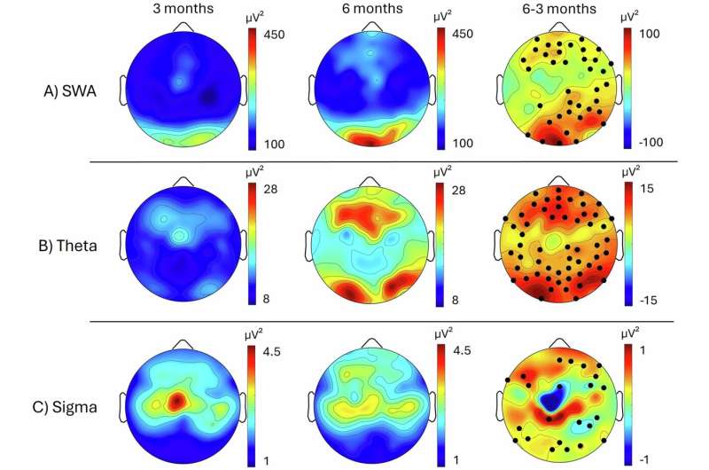
Topographical distribution of sleep EEG power. Credit:npj Biological Timing and Sleep(2026). DOI: 10.1038/s44323-026-00071-7
Electrical signals from the brain could help identify potential issues in the organ's development, a new study reports. Scientists from the University of Fribourg in Switzerland and the University of Surrey investigated electrical activity in the brains of sleeping infants longitudinally, at ages 3 and 6 months. They examined three electrical signals with distinct frequencies: slow wave activity (0.75–4.25 Hz), theta (4.5–7.5 Hz) power and sigma (9.75–14.75 Hz) power, which are key markers of sleep depth and brain development.
The researchers hope that their study, nowpublishedinnpj Biological Timing and Sleep, will enable better tracking of infants' neurodevelopment in the first months of life and reveal individual maps of brain maturation early on.
Dr. Salome Kurth, Research Group Leader at University of Fribourg, said, "At birth, the human brain size has reached only 27% of its adult size. Development happens rapidly in the first months of life, which is therefore a critical period for brain maturation. Sleep plays a central role in neuronal development; however, very little is known about the electrical signals the brain generates while an infant rests and the impact they have on their neurological maturation process.
"Using EEG (electroencephalography) is noninvasive and can provide us with an opportunity to map brain activity, track how it matures, and assess its influence on later behavioral outcomes."
To learn more,high-density EEGrecordings (during which a net with 124 sensors was placed on the scalp) were taken of 11 healthy infants during their regular sleep time. Behavioral development, focusing on gross motor skills and personal/social ability, of the infants was also assessed.
The research team found that between 3 and 6 months, electrical signal intensity changed across all frequencies. This likely reflects increases in the number ofsynaptic connectionsbetween neurons and increases in the wiring insulation through myelin, a fatty substance that wraps around nerve fibers and serves to increase the speed of electrical communication between neurons. These changes make visible how rapidly and where exactly the infant brain reorganizes, with shifting patterns of sleep-related brain activity reflecting the underlying maturation of neural networks.
Scientists note that continued monitoring ofsleep brain activitycould highlight issues with neuronal network growth early on with the potential for early diagnosis and intervention for a number of neuropsychiatric conditions such as ADHD.
The research team also found an association between the individual change in electrical signals during this period and behavioral development. It was identified that greater power increases over frontal regions of the brain correlated with more advanced skills at six months across behavioral domains. In particular, higherfrontal theta powerwas linked to better gross motor skills whereas higher frontal sigma power was associated with better personal social skills.
Dr. Valeria Jaramillo, a Wellcome Early Career Fellow at the University of Surrey, said, "Many neurodevelopmental disorders are not diagnosed until school age, highlighting the need to find a new way to identify such conditions early before symptoms emerge. Tracking sleep brain activity using EEG is a promising technique to spot when the brain has not matured as expected and to identify early indicators of developmental delays."
More information Matthieu Beaugrand et al, Tracing infant sleep neurophysiology longitudinally from 3 to 6 months: EEG insights into brain development, npj Biological Timing and Sleep (2026). DOI: 10.1038/s44323-026-00071-7


Post comments