
by Chris Casey,CU Anschutz Medical Campus
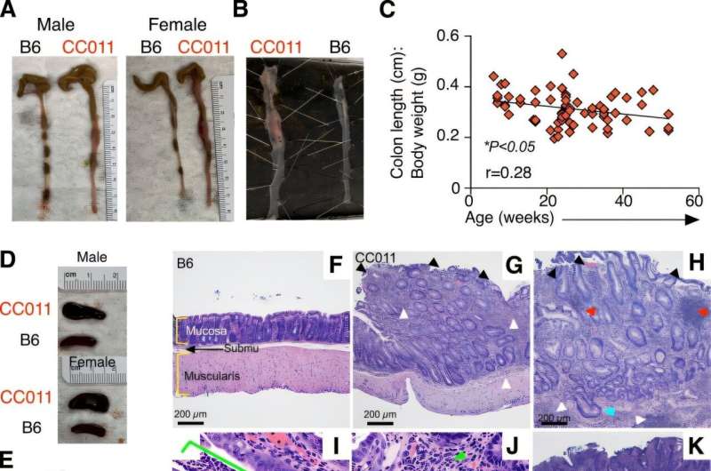
CC011 develop spontaneous colitis after 20 weeks of age with histological features that emulate human disease. Credit:Mucosal Immunology(2025). DOI: 10.1016/j.mucimm.2025.05.006
CU Anschutz researchers uncovered a link between a type of mucosal immune cell and gut inflammation, finding that the cells exacerbate inflammation and lead to chronic disease in certain conditions.
A new study yielded the potentially harmful effects of mucosal-associated invariant T cells (MAIT) on the intestinal barrier—the first such study to make this connection.
By genetically disrupting MAIT cells, researchers were able to halt ulcerative colitis in a strain of mice that are known for spontaneously developing chronic inflammation in the colon. The work ispublishedin the journalMucosal Immunology.
"We'd like to move forward to understand themolecular mechanismin the MAIT cells that is contributing to this," said Liyen Loh, Ph.D., the study's lead author and assistant research professor of immunology and microbiology at the CU Anschutz School of Medicine (SOM).
Researchers plan to involve human participants in future studies with the goal of developing novel therapeutics for inflammatory bowel diseases (IBD), including Crohn's disease and colitis.
Unlike other T cells, which recognize foreign-invader antigens in patient-specific fashion, MAIT cells recognize antigens in an identical manner across the population, said Laurent Gapin, Ph.D., professor of immunology and the study's lead author.
"That gives us an opportunity to come up with potential treatments that are going to be universal," he said. "There is a huge appeal not only in the IBD field but in autoimmunity and the cancer field as well to target the roles MAIT cells are potentially playing."
Several departments and divisions in the SOM, as well as collaborators in France, contributed to the study.
Loh and Gapin explain their research findings in the following interview, which has been edited for length and clarity.
Loh: They are unconventional T cells that respond at the mucosal surface of the intestines and other barrier sites where you would first sense inflammation and infection. They recognize metabolites (compounds produced by bacteria and other microorganisms) and they can sense when things are not right in the immune system.
They are potent for antimicrobial immunity, so that's their defense mechanism. But at the same time, they can also play a role in the inflammatory environment. So, when not kept under control, they can be activated in a harmful way—causing inflammation.
Gapin: Most T lymphocytes express receptors that detect foreign antigens in the form of peptides—small fragments of proteins—presented by HLA (histocompatibility leukocyte antigens), which are highly polymorphic across the human population. In contrast, MAIT cells, which can comprise up to 10% of T cells inhuman blood, recognize antigens that are not peptides.
Instead, their receptors detect small metabolite structures, including intermediates generated during the microbial synthesis of riboflavin. Although allhuman cellsrequire riboflavin, we cannot synthesize it ourselves and must rely on dietary sources and the metabolic activity of commensal bacteria in the gut.
These bacteria also produce intermediate metabolites in the riboflavin pathway, and it is one of these bacterial-derived intermediates that is specifically recognized by MAIT cell receptors. This enables MAIT cells to detect a wide range of bacteria and fungi with pathogenic potential.
Gapin: Out of those particular mice, we tested if there was an enrichment for bacteria that were making those particular compounds in their gut microbiota. The answer was yes. Compared to the control mice, there was an enrichment for bacteria that seems to produce those particular compounds.
So there is an enrichment taking place, leading to the possibility of having more antigens to stimulate MAIT cells. Also, a group in France is doing research to determine the genetic composition of this particular strain of mouse that leads to an increase in MAIT cells.
Loh: Along with the MAIT cells, there are other important T cells and innate cells that contribute to the disruption of the mucosa at the gut barrier. So together, they all contributed to the inflammation, but we couldn't quite pinpoint which came first. So, we genetically disrupted the T cell receptor locus to try and delete MAIT cells from these mice to understand if they weren't there, what happened? Did these mice go on to continue to develop colitis?
From our experiments where we did take out the MAIT cells, it seemed that they impacted inflammation. They had a very key role in the development of the disease, but, mechanistically, we couldn't delve any deeper to understand the precise mechanisms—whether they supported other innate cells to produce certain regulatory molecules such as cytokines.
Gapin: Our study was the first to describe the MAIT cells and colitis. We discovered if we prevent the development of MAIT cells in these mice, the colitis did not develop later on. But the reason why remains unclear.
Loh: In an inflammatory setting, you have disrupted intestinal barriers where, potentially, more microbial-derived metabolites are in contact with MAIT cells. This could be a mechanism by which the MAIT cells are being chronically stimulated.
Gapin: So, which comes first is not clear. Is it because you have a change in the microbiota that you have more MAIT cells, or is it because the genetics of this particular strain of mouse not only increase MAIT cells, but also change the microbiota? It's very complex. There is probably an interplay between genetics, microbiota and, indeed, maybe MAIT cells in the middle that are responding to both.
Loh: With humans, the limitation has been looking at the frequency of MAIT cells in patients' blood, and then further phenotypic studies in the tissues themselves. MAIT cells are thought to migrate to the site of inflammation where researchers see that they increase in numbers. But we really don't understand what these correlations mean.
That's why we wanted to look at an in vivo model that emulates the course of human disease, to see what was happening to MAIT cell numbers and their function. In future studies, we will try and tie the two—mouse model with human tissues—to understand how translationally relevant the model is to what's going on in patient tissues.
Loh: Yes, we think there are therapeutically relevant targets, so this study shows that it's worthwhile pursuing this mode. And it's not just for chronic intestinal inflammation.
In the cancer field, therapies (from this vein of research) are also being pursued. MAIT cells are universally relevant T cells that are present in every individual and highly conserved. They recognize specific targets that other T cells don't. So, they are ideal and novel players in therapeutics for IBD treatment.
Gapin: If MAIT cells contribute to disease pathology, blocking their activity could have therapeutic relevance, and there may be feasible strategies to achieve this. It is important to note that the system of antigen recognition by conventional T cells is highly patient-specific, shaped by the extreme polymorphism of HLA molecules across the human population.
MAIT cells, however, are fundamentally different. They employ a mode of antigen recognition that is essentially identical across individuals. In other words, while conventional T cell responses are highly variable between people, MAIT cells function in a uniform manner in both you and me.
This unique universality creates an opportunity to design therapeutic strategies that would be broadly applicable, independent of genetic background.
Such an approach is particularly compelling in contexts like inflammatory bowel disease (IBD), where blocking MAIT cell activity could provide a universal treatment avenue.
More information: Liyen Loh et al, MAIT cells exacerbate colonic inflammation in a genetically diverse murine model of spontaneous colitis, Mucosal Immunology (2025). DOI: 10.1016/j.mucimm.2025.05.006
Provided by CU Anschutz Medical Campus


Post comments