
Credit: Pixabay/CC0 Public Domain
For every motor skill you've ever learned, whether it's walking or watchmaking, there is a small ensemble of neurons in your brain that makes that movement happen. Our brains trigger these ensembles—what we sometimes call "muscle memories"—to get our bodies cooking, showering, typing, and every other voluntary thing we do.
Yet there's something missing from that picture: Despite years of research, no one is quite sure how the ensembles form in the first place.
Now, researchers led by Wu Tsai Neurosciences Institute affiliate Jun Ding are hoping to change that. Ding's team has taken important new steps toward understanding how our brains build the neural networks underlying motor memories.
The team's latest findings reveal that each network goes through dramatic changes as we learn a new skill. What begins as an almost random collection of neurons—with almost random links between them—evolves as our movements become more practiced and coordinated into a stable, efficient representation of that movement.
Those results, published in two papers—one in Cell Reports and one in Nature—could also inform a debate about how Parkinson's disease impairs movement. According to current thinking, that's because Parkinson's makes it difficult to activate existing muscle memories. But the new studies, taken together with previous evidence from Ding's lab and others, suggest the disease might instead destabilize the memories themselves.
That could also affect how doctors treat Parkinson's.
"Often we try to reactivate motor memories with a drug like L-Dopa, but if the memory actually is disrupted, that won't work, " said Ding, an associate professor of neurosurgery and of neurology and neurological sciences at Stanford Medicine. "So we actually need to think about what's the most efficient way to reactivate the system and regain the ability to learn."
Learning to run
In a set of experiments, the lab focused on neurons in the striatum, a part of the brain that helps coordinate movements and learn new ones.
The striatum is also home to our so-called muscle memories, the neural ensembles that encode different movements.
To probe how these ensembles form as we learn a new skill, postdoctoral fellow Omar Jáidar, graduate student Eddy Albarran, and collaborators set mice atop a spinning wheel. Then, they watched as those mice learned to run in this novel and somewhat precarious position during short sessions over the course of about a week. All the while, they tracked neural activity in the striatum with two-photon fluorescence imaging.
When the mice first hopped on the wheel, the striatum was as uncoordinated as the mice were: About three-quarters of the neurons the team could see fired, seemingly at random. But as mice got more coordinated, so too did activity in the striatum, which consolidated into a smaller, more consistent network. A muscle memory was born.
Zooming in
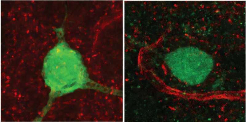
In another experiment, Ding's postdoctoral fellow Mengjun Sheng and colleagues showed that something similar happens to the synaptic links connecting neurons in the primary motor cortex to the striatum.
Researchers presume that the primary motor cortex, which is responsible for planning movements, plays a key role in building and refining the muscle memories in the striatum as a movement becomes more practiced. But again, how this happens has been unclear.
To investigate how learning a new skill changes the wiring between cortex and striatum, Sheng and colleagues followed mice as they learned to press a lever to get a sip of water. Using two-photon imaging, they tracked thousands of synapses linking specific neurons in the primary motor cortex to the striatum.
"What we found challenges the conventional view, " Ding said.
Researchers had long thought that when a neuron fired, each of its synapses would transmit the same signal to other neurons.
That was not the case. When mice were first learning the lever-pressing task, synapses sent mixed signals: When a motor cortex neuron fired, some of its synapses in the striatum passed the signal along, some didn't, and the pattern would change from one lever press to the next.
But as with the ensembles encoding movements, the set of synapses connecting each motor cortex neuron to the striatum started out large and uncoordinated, but grew smaller, more efficient, and more coordinated over time.
Ding and colleagues say their data reveal in new detail how the brain reorganizes itself—even at the level of axons—to more efficiently encode and drive movements. As mice figure out how to press a lever or run on a wheel, their brains are refining from top to bottom the circuits that make those movements possible.
New ways to look at Parkinson's disease
The study adds to our understanding of how skills like driving, gardening, or playing an instrument tend to become more efficient and reliable with time—but it may also help inform a debate about how Parkinson's disease works.
According to one standard viewpoint, Parkinson's disease works by interfering with the brain's ability to activate motor memories, while the memories themselves remain intact.
Ding's research, on the other hand, suggests something else is going on. In a previous study, he and colleagues found that when mice were given a drug that simulated the effects of Parkinson's, the mice lost many of their striatal synapses. At the same time, they also grew new synapses, Ding said.
Those findings hint that Parkinson's doesn't simply eat away at our ability to activate motor memories. Instead, it might destabilize established muscle memory patterns, sending them back into the more uncoordinated state seen prior to motor learning.
"We don't have the direct evidence yet, " to say whether that's exactly what happens in Parkinson's, Ding said. For that, he and his lab will need to teach mice new skills, then see if Parkinson's-simulating drugs break the synapses underlying those muscle memories.
But if Ding's ideas are correct, treatments that stabilize synapses could stave off motor memory loss. At the same time, doctors might be able to use L-Dopa—which transforms into dopamine, a "teaching molecule" that helps the brain learn—specifically during physical therapy to help patients recover some of what they've lost.
"These aren't precise predictions, but my thoughts are along those lines, " Ding said. "The next step is to look at mouse models of Parkinson's and see what we can learn."
More information: Omar Jáidar et al, Refinement of efficient encodings of movement in the dorsolateral striatum throughout learning, Cell Reports (2025). DOI: 10.1016/j.celrep.2025.116229 Mengjun Sheng et al, Remodelling of corticostriatal axonal boutons during motor learning, Nature (2025). DOI: 10.1038/s41586-025-09336-w Journal information: Cell Reports, Nature
Mengjun Sheng et al, Remodelling of corticostriatal axonal boutons during motor learning, Nature (2025). DOI: 10.1038/s41586-025-09336-w


Post comments