
byOhio State University Medical Center
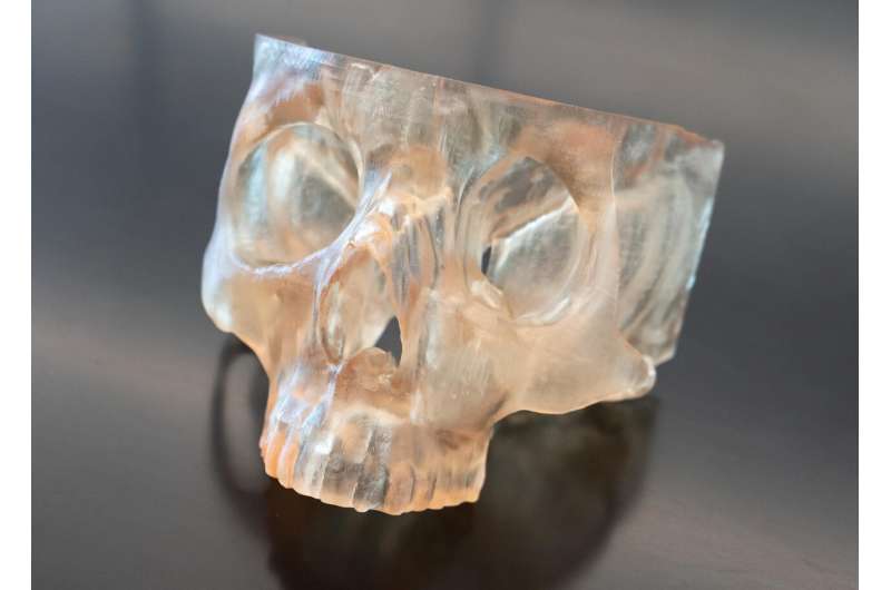
This 3D model of a patient's jaw was created by doctors at The Ohio State University Comprehensive Cancer Center–Arthur G. James Cancer Hospital and Richard J. Solove Research Institute in Columbus, OH. The 3D model helps surgeons precisely locate where the cancer is inside the patient's jaw. Credit: The Ohio State University Comprehensive Cancer Center
Using 3D modeling to plan and guide cancer surgeries increases surgical precision, resulting in complete tumor removal for 92% of head and neck cancers that have invaded bone, according to a new study published by The Ohio State University Comprehensive Cancer Center–Arthur G. James Cancer Hospital and Richard J. Solove Research Institute (OSUCCC–James).
The research ispublishedin the journalOral Oncology.
"The precision of what we take out is critical to ensure we get the whole tumor, but not so much that we're devastating the patient's function in the long term and taking out things that don't need to be removed," said Kyle VanKoevering, MD, an otolaryngologist (head and neck surgeon) at the OSUCCC–James and medical director of the M4 Lab (Medical Modeling, Materials and Manufacturing Lab) within the Ohio State College of Engineering. "This 3D modeling being completely personalized to each patient is really helping improve the precision in the operating room."
For this research, VanKoevering and team compared the surgical outcomes of 68 patients with bone-invading head and neck cancers treated at the OSUCCC–James. Thirty-seven patients received in-house 3D models for intraoperative use while the other 31 did not. The study population was predominantly male, and nearly all were active or previous tobacco users (94.6%).
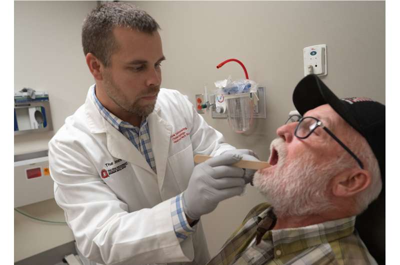
Kyle VanKoevering, MD (left) assesses healing of the jaw he rebuilt for oral cancer survivor, Greg McAlarney (right). Credit: The Ohio State University Comprehensive Cancer Center
Surgeries with access to apatient-specific 3D modelto use as a visual planning aid in the operating room had better negative surgical margins—meaning the surrounding tissue showed no evidence of cancer—compared with the group that did not have the assistance of a 3D model.
"This model is especially critical incancers that have invaded bone, because tumor boundaries are often less visible or palpable. Our 3D models are built based on the patient's actual tumor imaging, so it gives us a much better visual map at the patient's bedside for removing the cancer as completely as possible while also sparing important structures and tissue to maintain function after surgery," VanKoevering explained.
This study looked at the cancer-control impact of usingcustom, 3D-printed modelsof a patient's anatomy to plan complex surgeries impacting the head and neck, where delicate structures that impact speech, chewing and swallowing are often impacted.
Matthew Marquardt, study corresponding author and third-year medical student, noted that this is thefirst studyto evaluate the ability of 3D modeling to help improve cancer control in the operating room.
"This really sets the stage for larger studies looking at how 3D modeling can enhance surgery planning and precision, not just in the field of head and neck cancer surgery but in other areas that involve bone and soft tissue, like orthopedics," said Marquardt, who worked on this project as part of a focused medical school research year through the Pelotonia Scholars Program.
"Long term, our hope is that this work will enable other surgeons to use this technology across the country to help improve people's lives and improve cancer outcomes," said Marquardt.
More information Matthew D. Marquardt et al, In-house 3D modeling associated with margin-negative resection in mandibular oral cavity malignancies, Oral Oncology (2025). DOI: 10.1016/j.oraloncology.2025.107588
Provided by Ohio State University Medical Center


Post comments