
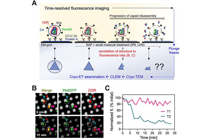
An in vitro assay to study HIV-1 uncoating by CLEM. Credit: ACS Nano (2025). DOI: 10.1021/acsnano.5c06724
40 million people live with HIV globally, and that number continues to rise. While therapies exist to reduce the amount of HIV in a patient's body and, in turn, reduce HIV symptoms, there remains no cure. Engineering better drugs and eventually a cure depends on our ability to answer foundational questions like: How does HIV invade and replicate in host cells?
Salk Institute scientists, led by Associate Professor Dmitry Lyumkis and graduate student researcher Zaida Rodriguez, developed an improved workflow for characterizing capsids (genome-containing protein cones) in HIV-1 (the most common form of HIV). HIV-1 capsids protect the viral genome and enable host cell invasion. The new workflow uses correlative light and cryo-electron microscopy techniques to capture the morphological changes of HIV-1 capsids over time. The findings were published in ACS Nano on August 22, 2025.
In 2024, the drug lenacapavir, which blocks viral replication by altering the HIV-1 capsid structure, was deemed the breakthrough of the year by Science magazine. Still, structural studies of how capsids form, disassemble, and stabilize have been restricted by workflow and technology limitations.
This novel workflow incorporates advanced technology that will allow scientists to better understand how capsid morphology changes in response to distinct pressures and to pinpoint new, more effective ways to target capsids and treat HIV-1. Importantly, the workflow can also be used to study structural changes in other, non-HIV viruses.
More information: Zaida K. Rodriguez et al, Time-Resolved Fluorescence Imaging and Correlative Cryo-Electron Tomography to Study Structural Changes of the HIV-1 Capsid, ACS Nano (2025). DOI: 10.1021/acsnano.5c06724 Journal information: ACS Nano, Science


Post comments