
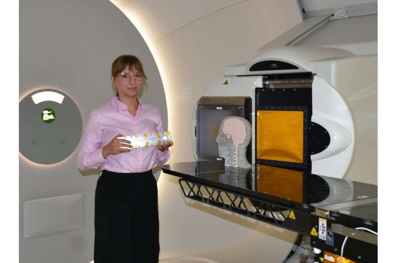
The irradiation station at the Bronowice Cyclotron Center. Ph.D. student Paulina Stasica-Dudek from the Institute of Nuclear Physics of the Polish Academy of Sciences presents the detector used to measure the quality of the proton beam. Credit: IFJ PAN
Modern methods of radiotherapy would fight cancer more effectively and safely if treatments could be planned, taking into account the radiation quality of the therapeutic proton beams. An achievement by physicists from the Institute of Nuclear Physics of the Polish Academy of Sciences in Cracow brings us closer to this goal. The research is published in the journal Physics in Medicine & Biology.
In today's medicine, the relationship is simple: if we want cancer treatment to be more effective and safer for the patient, we usually have to invest in better irradiation equipment, which, in the case of modern proton beams, means costs running into tens of millions of euros.
It turns out, however, that an improvement in treatment is achievable at a relatively low cost using the equipment already in operation, thanks to a solution presented by a team of physicists from the Cyclotron Center Bronowice (CCB) of the Institute of Nuclear Physics of the Polish Academy of Sciences (IFJ PAN) in Cracow. The innovation, which can be applied with the equipment already in use, allows medical physicists, at the therapy planning stage, to verify a parameter not previously exploited in clinical practice: the quality of the proton beam.
"When we take the quality of the radiation beam into account, we are able to determine the biological effect of radiation more precisely and destroy cancer cells more effectively, while reducing damage to healthy tissues. Modern proton therapy planning software allows this parameter to be taken into account.
"In practice, nobody does it, and one of the reasons for this is that until now, there has been no quick and simple method for experimental verification of radiation quality. We have shown that there is a practical solution to this problem that is cheap and easy to implement, " says Dr. Jan Gajewski (IFJ PAN).
In the fight against cancer cells, doctors currently use irradiation mainly with photons, while only a small number of patients undergoing radiotherapy can benefit from proton therapy. Two facts are key here. Firstly, when a beam of high-energy particles interacts with the cellular DNA molecules, it damages them, eventually causing cell death. Secondly, cancer cells are generally more sensitive to radiation than healthy, normal tissue cells that can efficiently repair DNA damage.
How the therapeutic beam is administered becomes critical. To avoid later complications, such as necrosis of healthy tissue, the beam should interact with cells within the tumor only. This is, however, not possible, because radiation must typically cross normal tissue to reach the tumor.
At the same time, the entire volume of the tumor with some surrounding healthy tissue must be irradiated safely. This is because, in practice, if some of the cancer cells survive the treatment, the recurrence is often more dangerous than the primary tumor.
All radiotherapy treatments are carefully planned. The physician first determines the extent of the cancerous lesion and then, using specialized software, plans a series of treatment fractions typically spanning over weeks.
Physicians and physicists together precisely determine the directions from which the tumor will be irradiated, the energies of the particles to be used, the intensity of the beam, as well as irradiation duration. Appropriate treatment planning later makes it possible to deliver a lethal dose of radiation to the tumor volume while minimizing damage to healthy tissue.
However, planning alone is not enough. In order to confirm that the therapeutic dose conforms with the developed treatment plan, before each treatment the medical physicist must measure the distribution of radiation doses the patient will receive.
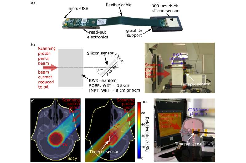
(a) The MiniPIX Timepix3 detector with 300 µm-thick silicon sensor in Flex configuration (Advacam, Prague). Adapted from Stasica-Dudek et al (2025). CC BY 4.0. (b) A scheme (left) and a photo (right) of the experimental setup for the SOBP and IMPT field measurements behind a solid RW3 phantom. (c) IMPT treatment plan prepared for the CIRS head phantom (left), one of the fields recalculated for half of the head with indicated sensor position (middle), and a photo of the experimental setup for the measurements behind a heterogeneous phantom (right). The sensor thickness in the middle panel is not to scale. Credit: Physics in Medicine & Biology (2025). DOI: 10.1088/1361-6560/adcaf9
If the therapeutic beam consists of photons, they all interact similarly with the cells encountered in the patient body. When it comes to the biological effects of irradiation, physicists talk about the quality of the radiation, which for photons will be the same at each irradiated point. Protons behave differently when slowing down: initially, they lose little energy, but the slower they are, the more rapidly they lose energy.
As a result, the proton beam deposits most of its energy at the end of its path, at a well-defined depth inside the patient's body. The quality of such a proton beam is significantly different from that of a photon beam commonly used in radiation therapy.
A physical parameter called linear energy transfer (LET) is used to describe the quality of radiation that influences the biological effect. It carries information about how much energy a particle deposits over a fixed distance along its path and how this loss varies with the energy of the particle.
Preparing treatment plans taking into account the LET parameter is not a problem today. Unfortunately, current clinical practice lacks instruments and measurement techniques to verify the distribution of linear energy transfer directly in the clinical environment of radiotherapy.
During research carried out at CCB using a proton beam from the Proteus C-235 cyclotron, scientists from IFJ PAN used a commercially available Timepix3 detector to characterize the LET parameter. This small and relatively easy-to-use detector is a result of the work of the Medipix3 Collaboration, formed in 2005 at the European Organization for Nuclear Research (CERN) with a view to broad applications of technical solutions developed for particle tracking in high-energy physics experiments.
The numerous applications of the Timepix detectors are an exemplary demonstration of how pioneering physics research, seemingly disconnected from everyday reality, translates into improvements in the quality of human life.
"The Timepix3 detector is equipped with a 300 micrometer thick silicon sensor. Its readout electronics is a 256 by 256 pixel matrix, which allows the acquisition of single particle tracks. The response of each pixel depends on the energy it registers.
"This information, together with the track parameters, allows—using artificial intelligence methods—the identification of a single proton and the estimation of its LET parameter. The proposed methodology makes it possible to characterize LET under irradiation conditions compatible with therapeutic plans, which was a key challenge for us, " explains doctoral student Paulina Stasica-Dudek (IFJ PAN), first author of the article.
Before the method for measuring the quality of proton beams proposed by the Cracow physicists becomes everyday practice, a technical hurdle will have to be overcome. Modern medical cyclotrons produce beams for clinical use, i.e., of high intensity.
Meanwhile, measurements carried out with Timepix detectors require low-intensity beams. It is known, however, that this problem can be solved by cyclotron manufacturers with an appropriate update to the software controlling the intensity of the beam produced by a cyclotron.
"For the first time, we can speak of a practically ready-to-implement method of measuring the quality of the radiation beam directly in proton therapy facilities. Its dissemination is a way to improve the efficiency and safety of modern proton therapy and more advanced irradiation methods using helium, carbon or oxygen ion beams, " says Dr. Antoni Ruciński, professor at IFJ PAN, summarizing the work of the team.
Research on the methodology for measuring the quality of radiation beams was carried out under the LIDER XII grant from the National Centre for Research and Development (Poland).
More information: Paulina Stasica-Dudek et al, Experimental validation of LET in intensity-modulated proton therapy with a miniaturized pixel detector, Physics in Medicine & Biology (2025). DOI: 10.1088/1361-6560/adcaf9 Journal information: Physics in Medicine and Biology


Post comments