

Introduction
Chest X-rays remain one of the most widely used diagnostic imaging tools globally, essential for detecting a broad range of thoracic conditions such as tuberculosis, pneumonia, and heart failure. With over 2 billion chest radiographs performed each year, they are a cornerstone of frontline diagnostics. However, interpretation challenges persist. Diagnostic accuracy can vary significantly among physicians due to factors like subjective judgment, fatigue, and limited radiological expertise—issues that are particularly pronounced in resource-constrained settings facing severe radiologist shortages. These limitations can lead to missed or delayed diagnoses, directly compromising patient outcomes.
In this context, open-source artificial intelligence (AI) models have emerged as transformative tools. Trained on large, diverse, multinational datasets and validated across varied clinical settings, these models serve as scalable digital assistants that enhance chest X-ray interpretation. Studies show that AI-assisted interpretation not only improves diagnostic accuracy and sensitivity but also accelerates workflow and reduces cognitive burden—allowing even non-radiologists to achieve near-expert performance. Crucially, open-access AI platforms such as Ark+ and X-Raydar are proving to be as accurate as proprietary systems, while offering free, adaptable solutions for deployment in low-resource environments. By democratizing access to high-quality diagnostic support, open-source AI has the potential to bridge critical global health gaps, transforming care delivery for underserved populations.1,2,3,4
Enter AI: Redefining Chest X-Ray Analysis
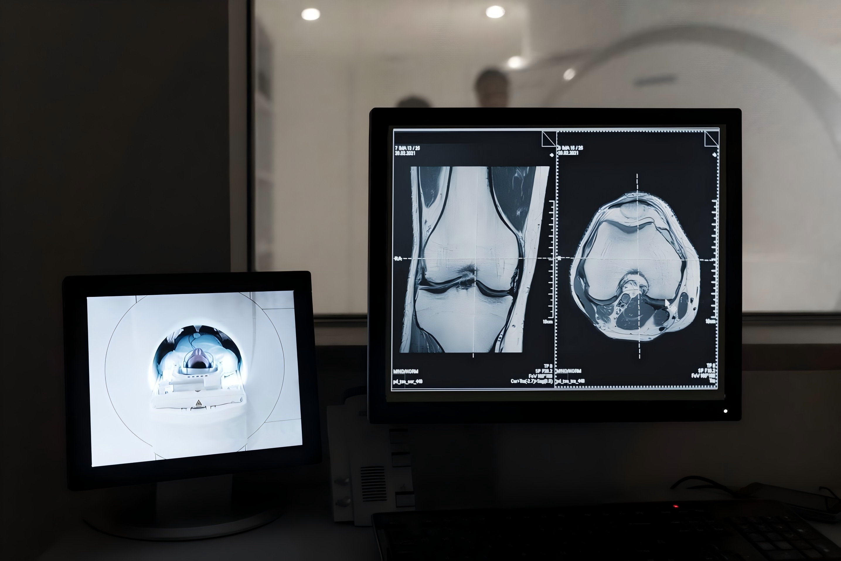
The integration of artificial intelligence into chest X-ray interpretation is redefining diagnostic possibilities. At the core of this transformation are deep learning models—particularly convolutional neural networks (CNNs)—trained on large, publicly available datasets such as NIH’s ChestX-ray14, Stanford’s CheXpert, and MIT’s MIMIC-CXR. These datasets contain hundreds of thousands of annotated images labeled for a wide spectrum of thoracic abnormalities.
Modern AI systems can now accurately classify and detect pathologies such as consolidation, pneumothorax, cardiomegaly, and pleural effusion. Notably, these models often match or exceed the diagnostic performance of general radiologists in specific clinical tasks. Multi-center validation studies confirm their robustness across diverse patient populations and imaging conditions. For instance, a 2024 FDA-cleared AI system demonstrated improved diagnostic accuracy and efficiency for both radiologists and non-specialists when used as a decision-support tool.
Rather than replacing human expertise, AI functions as a clinical partner—enhancing diagnostic precision, streamlining workflows, and providing expert-level support in settings where trained radiologists may be unavailable. This collaborative approach is already reducing diagnostic errors and enabling more timely, effective clinical decision-making.5,6,7,8,9
Real-World Applications and Impact
The real-world application of open-source AI in chest X-ray interpretation is yielding substantial benefits, particularly in regions with limited access to radiological services. These tools empower frontline clinicians—especially in rural or low-resource settings—by providing rapid, reliable image analysis where expert interpretation is often unavailable. By acting as a second reader, AI improves diagnostic accuracy for non-specialists and supports the early identification of critical conditions, facilitating timely triage and intervention.
Beyond primary care clinics, AI-enabled chest X-ray analysis is being integrated into a range of clinical environments, including emergency departments, mobile imaging units, and humanitarian response settings. In these contexts, FDA-cleared AI systems have demonstrated reductions in image interpretation time and parity with radiologists in diagnostic performance, validating their utility in high-pressure, real-time clinical workflows.
Open-source frameworks further enhance accessibility by offering free, customizable solutions that can be adapted to local needs. These models not only support widespread adoption but also foster continuous improvement through global collaboration. Clinical studies underscore their role in improving detection rates for diseases like tuberculosis, alleviating pressure on overstretched healthcare systems, and expanding diagnostic capacity across medical specialties.
However, ongoing clinical validation and continued training on diverse, real-world datasets remain essential to ensure reliability and minimize bias. Addressing these challenges will be critical to realizing the full potential of AI in global health.10,11,12
Reference:
Anderson, P.G., Tarder-Stoll, H., Alpaslan, M. et al. Deep learning improves physician accuracy in the comprehensive detection of abnormalities on chest X-rays. Sci Rep 14, 25151 (2024). https://doi.org/10.1038/s41598-024-76608-2
López Alcolea J, Fernández Alfonso A, Cano Alonso R, Álvarez Vázquez A, Díaz Moreno A, García Castellanos D, Sanabria Greciano L, Hayoun C, Recio Rodríguez M, Andreu Vázquez C, Thuissard Vasallo IJ, Martínez de Vega V. Diagnostic Performance of Artificial Intelligence in Chest Radiographs Referred from the Emergency Department. Diagnostics (Basel). 2024 Nov 18;14(22):2592. doi: 10.3390/diagnostics14222592. PMID: 39594258; PMCID: PMC11592727.
Lee KH, Lee RW, Kwon YE. Validation of a Deep Learning Chest X-ray Interpretation Model: Integrating Large-Scale AI and Large Language Models for Comparative Analysis with ChatGPT. Diagnostics (Basel). 2023 Dec 30;14(1):90. doi: 10.3390/diagnostics14010090. PMID: 38201398; PMCID: PMC10795741.
Vijayan S, Jondhale V, Pande T, Khan A, Brouwer M, Hegde A, Gandhi R, Roddawar V, Jichkar S, Kadu A, Bharaswadkar S, Sharma M, Vasquez NA, Richardson L, Robert D, Pawar S. Implementing a chest X-ray artificial intelligence tool to enhance tuberculosis screening in India: Lessons learned. PLOS Digit Health. 2023 Dec 7;2(12):e0000404. doi: 10.1371/journal.pdig.0000404. PMID: 38060461; PMCID: PMC10703224.
Anand A, Krithivasan S, Roy K. RoMIA: a framework for creating Robust Medical Imaging AI models for chest radiographs. Front Radiol. 2024 Jan 8;3:1274273. doi: 10.3389/fradi.2023.1274273. PMID: 38260820; PMCID: PMC10800823.
Chen Y, Wan Y, Pan F. Enhancing Multi-disease Diagnosis of Chest X-rays with Advanced Deep-learning Networks in Real-world Data. J Digit Imaging. 2023 Aug;36(4):1332-1347. doi: 10.1007/s10278-023-00801-4. Epub 2023 Mar 29. PMID: 36988837; PMCID: PMC10054207.
Johnson, A.E.W., Pollard, T.J., Berkowitz, S.J. et al. MIMIC-CXR, a de-identified publicly available database of chest radiographs with free-text reports. Sci Data 6, 317 (2019). https://doi.org/10.1038/s41597-019-0322-0
López Alcolea J, Fernández Alfonso A, Cano Alonso R, Álvarez Vázquez A, Díaz Moreno A, García Castellanos D, Sanabria Greciano L, Hayoun C, Recio Rodríguez M, Andreu Vázquez C, Thuissard Vasallo IJ, Martínez de Vega V. Diagnostic Performance of Artificial Intelligence in Chest Radiographs Referred from the Emergency Department. Diagnostics (Basel). 2024 Nov 18;14(22):2592. doi: 10.3390/diagnostics14222592. PMID: 39594258; PMCID: PMC11592727.
Lee S, Kim EK, Han K, Ryu L, Lee EH, Shin HJ. Factors for increasing positive predictive value of pneumothorax detection on chest radiographs using artificial intelligence. Sci Rep. 2024 Aug 23;14(1):19624. doi: 10.1038/s41598-024-70780-1. PMID: 39179744; PMCID: PMC11343866.
Tseng WC, Wang YC, Chen WC, Lin KP. Development of an AI model for pneumothorax imaging: Dataset and model optimization strategies for real-world deployment. Eur J Radiol Open. 2025 Jun 10;14:100664. doi: 10.1016/j.ejro.2025.100664. PMID: 40547323; PMCID: PMC12179720.
de Camargo TFO, Ribeiro GAS, da Silva MCB, da Silva LO, Torres PPTeS, Rodrigues DdSdS, de Santos MON, Filho WS, Rosa MEE, Novaes MdA, Massarutto TA, Junior OL, Yanata E, Reis MRdC, Szarf G, Netto PVS and de Paiva JPQ (2025) Clinical validation of an artificial intelligence algorithm for classifying tuberculosis and pulmonary findings in chest radiographs. Front. Artif. Intell. 8:1512910. doi: 10.3389/frai.2025.1512910
Development and validation of open-source deep neural networks for comprehensive chest x-ray reading: a retrospective, multicentre study. Cid, Yashin Dicente et al. The Lancet Digital Health, Volume 6, Issue 1, e44 - e57


Post comments