
Integration of proteomics, transcriptomics and AI in identification of aging markers
Two recent studies published in Nature and Cell have utilized innovative technologies such as single-cell RNA sequencing, proteomics and machine learning models to study aging and disease progression, offering new insights into the biological basis for diagnostic principles of aging and age-related diseases. The work published in Nature introduces a new method to estimate the aging of major human organs using plasma proteins. The authors Oh et al. hypothesized that since specific organ-related proteins can be measured in plasma, it is possible to track the aging process of different organs using these plasma proteins. To test this hypothesis, they measured 4,979 proteins in the plasma of 5,676 individuals from five independent cohorts. They mapped these proteins to their putative organ sources using data from human organ bulk RNA sequencing. Then, they trained machine learning models to predict the aging of 11 major organs (adipose tissue, artery, brain, heart, immune tissue, intestine, kidney, liver, lung, muscle and pancreas) using the organ-enriched proteins as input.
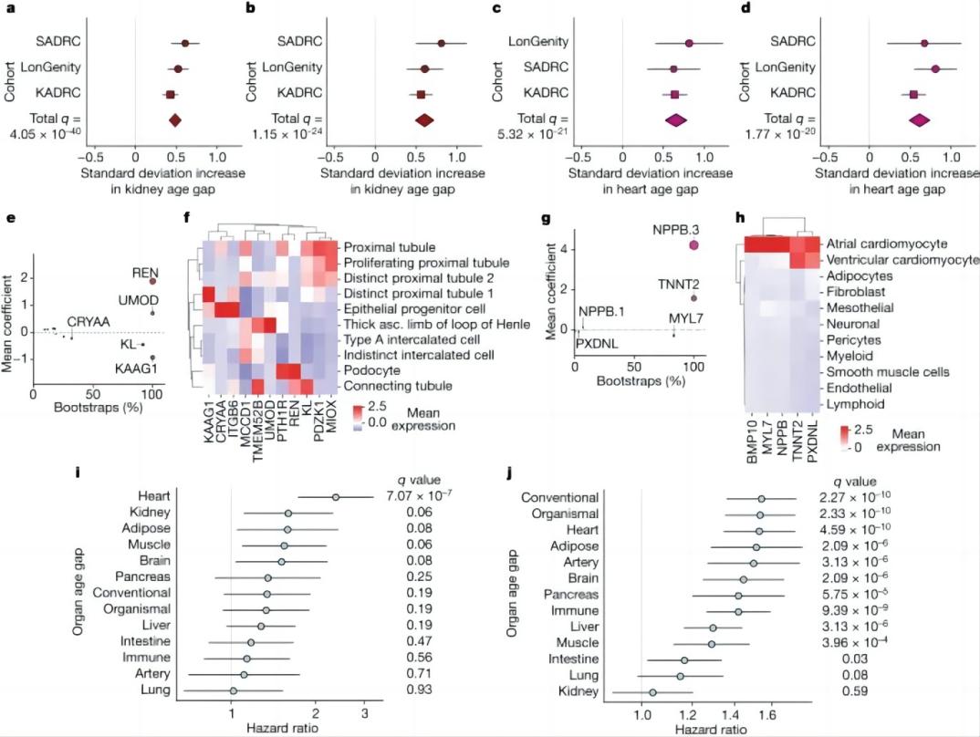
a, A cross-cohort meta-analysis of the association (linear regression) between the kidney age gap and hypertension (with hypertension n = 1,566, without n = 1,561). False discovery rate (FDR) P valuemeta = 4.05 × 10−40, effect sizemeta = 0.486. (Supplementary Table 10). b, As in a, kidney age gap versus diabetes (with diabetes n = 335, without n = 2,839). FDR P valuemeta = 1.15 × 10⁻²⁴, effect sizemeta = 0.604. c, As in a, heart age gap versus atrial fibrillation or pacemaker (with atrial fibrillation n = 239, without n = 2,936). FDR P valuemeta = 5.32 × 10⁻²¹, effect sizemeta = 0.657. d, As in a, but for heart age gap versus heart attack (with heart attack history n = 280, without n = 2,904). FDR P valuemeta = 1.77 × 10⁻²⁰, effect sizemeta = 0.615. e, All kidney aging model coefficients. x axis shows % of model instances in the bagged ensemble that include the protein. Size of bubbles is scaled by the absolute value of the mean model weight across model instances (absolute value of y axis) (Supplementary Table 7). f, Single-cell RNA expression of kidney51 aging model proteins. Mean normalized expression values shown. g, As in e, but for the heart aging model. h, Human heart single-cell RNA expression of heart52. Mean normalized expression values shown. i, Cox proportional hazard regression analysis of the relationship between organ age gap and future congestive heart failure risk over 15 years of follow-up in the LonGenity cohort for those without heart failure history at baseline (n = 26 events in 812 individuals). FDR P valueHeart = 7.07 × 10⁻⁷, hazard ratioHeart = 2.37. (Supplementary Table 11). j, Cox proportional hazard regression analysis of the relationship between organ age gap and future mortality risk, over 15 years of follow-up in the LonGenity cohort (n = 173 events in 864 individuals). FDR P valueConventional = 2.27 × 10⁻¹⁰, hazard ratioConventional = 1.54. (Supplementary Table 12). All error bars represent 95% confidence intervals.
Organ age predicts health and disease.Credit:DOI:https://doi.org/10.1038/s41586-023-06802-1
The models were trained in a cohort of 1,398 healthy individuals and then tested in four fully independent cohorts and in held-out test participants with dementia. The results showed that the models significantly estimated age in all five cohorts. Importantly, the models were able to identify individuals with accelerated aging in one or more organs. Approximately 18.4% of individuals had accelerated aging in only one organ, while 1.7% showed accelerated aging in multiple organs.
The authors further investigated the relationship between organ age and age-related diseases and found that accelerated organ aging was associated with higher mortality risk and increased risk of specific diseases. For example, individuals with accelerated heart aging had a 250% increased risk of heart failure. Additionally, accelerated brain and vascular aging independently predicted the progression of Alzheimer's disease.
A new method called FIBA (Feature Importance for Biological Aging) was also developed in this work to identify which proteins in the models were most important for predicting cognitive decline and Alzheimer's disease. Using this method, they identified a subset of brain aging proteins that had the strongest association with cognitive decline, such as Aldolase Fructose-Bisphosphate C (ALDOC), neuronal pentraxin receptor (NPTXR), carnosine dipeptidase 1 (CNDP1) and Lanc Like Glutathione S-Transferase 1 (LANCL1).
In conclusion, this study demonstrates that plasma proteomics and machine learning can be used to non-invasively measure organ health and aging in living individuals. The resulting organ aging models can predict mortality, organ-specific functional decline, disease risk and progression, and aging heterogeneity between tissues. This approach could potentially be used to understand the effects of health interventions at the organ level and to guide personalized medicine approaches to aging and age-related diseases.
Similarly, another work published in Cell introduces a novel approach, named TEMPO (tracing expression of multiple protein origins), which combines high-resolution liquid biopsy proteomics with single-cell transcriptomics and artificial intelligence (AI). Instead of analyzing proteomics data from plasma samples which reflect the multi-organ aging, the method developed by Wolf et al. is used to trace the cellular origin of proteins within fluid samples obtained from the anterior chamber and vitreous of the human eye.
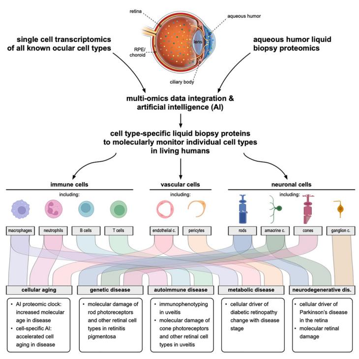
Credit:DOI:https://doi.org/10.1016/j.cell.2023.09.012
The authors analyzed 5,953 proteins detected in aqueous humor from 120 patients undergoing cataract surgery or vitrectomy. To identify the cell types expressing the genes for these proteins, they integrated proteomics data with single-cell RNA sequencing data from 82,072 cells from all known ocular and extra-ocular cell types. This analysis revealed 1,920 cell-type specific marker proteins.
Using TEMPO, the authors monitored molecular changes in retinitis pigmentosa, diabetic retinopathy, and uveitis patients, identifying cell types affected by these diseases. They also detected molecular changes in Parkinson's disease patients' aqueous humor, identifying retinal neurons as the main affected cell type.
Furthermore, they found that proteins from the eye's aqueous humor could be used to assess its molecular age using AI models. Surprisingly, these models predicted accelerated aging in several eye diseases not primarily related to chronological age.
The authors conclude that TEMPO represents a powerful tool to identify the contribution of specific cells to human disease and suggests that diseases may impact aging in distinct ways. The approach could transform molecular diagnostics and prognostics while uncovering new cellular disease and aging mechanisms.
Oh HS, Rutledge J, Nachun D, Pálovics R, Abiose O, Moran-Losada P, Channappa D, Urey DY, Kim K, Sung YJ, Wang L, Timsina J, Western D, Liu M, Kohlfeld P, Budde J, Wilson EN, Guen Y, Maurer TM, Haney M, Yang AC, He Z, Greicius MD, Andreasson KI, Sathyan S, Weiss EF, Milman S, Barzilai N, Cruchaga C, Wagner AD, Mormino E, Lehallier B, Henderson VW, Longo FM, Montgomery SB, Wyss-Coray T. Organ aging signatures in the plasma proteome track health and disease. Nature. 2023 Dec;624(7990):164-172.
Wolf J, Rasmussen DK, Sun YJ, Vu JT, Wang E, Espinosa C, Bigini F, Chang RT, Montague AA, Tang PH, Mruthyunjaya P, Aghaeepour N, Dufour A, Bassuk AG, Mahajan VB. Liquid-biopsy proteomics combined with AI identifies cellular drivers of eye aging and disease in vivo. Cell. 2023 Oct 26;186(22):4868-4884.e12.


Post comments